



Project Objectives
- Development of a prototype and utility model of a next-generation medical device for imaging the thyroid gland and other small organs in nuclear medicine, with a particular focus on radionuclide therapy and diagnostics.
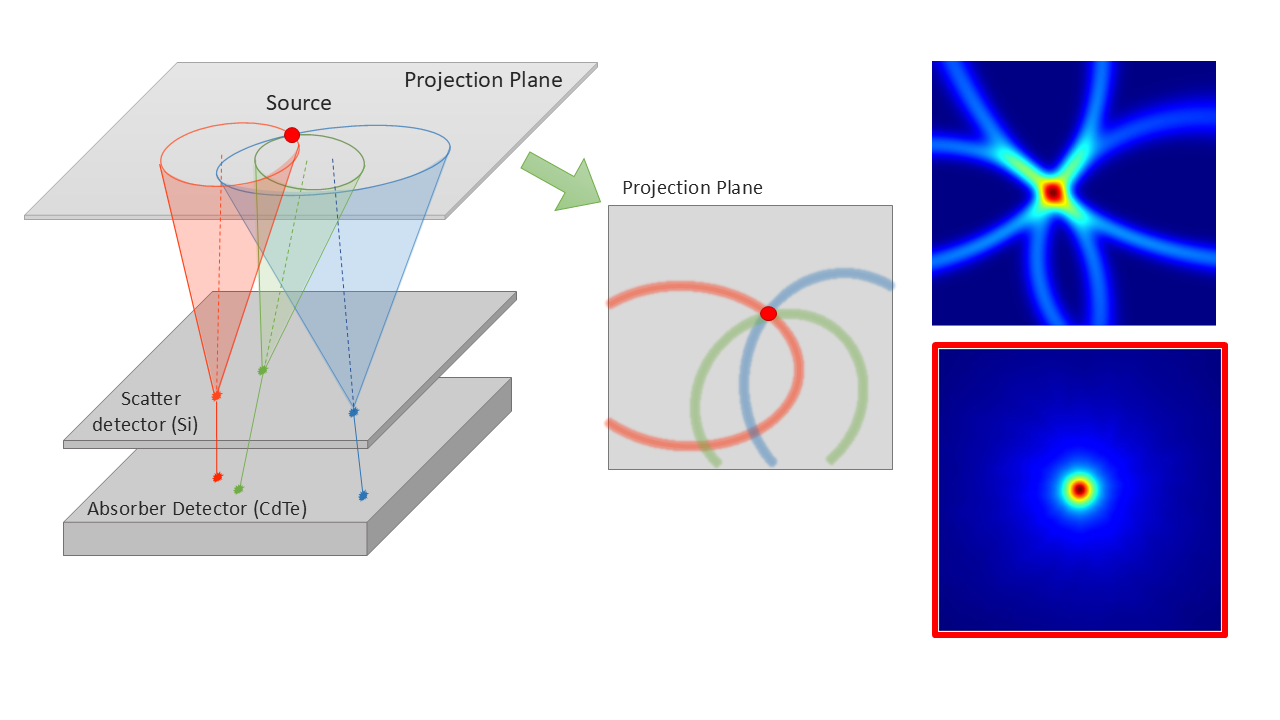
- Development of a novel detector in a Compton camera configuration based on Timepix3 technology, together with research and development of a new generation of detectors based on Timepix4 technology.
- Development of user software integrating robotic arm motion control, data acquisition, image reconstruction, data analysis, and communication with NIS/RIS/PACS systems using the DICOM 3.0 standard.
Project Strategy
The consortium is developing an advanced imaging system designed primarily for monitoring the thyroid gland throughout diagnosis and radioiodine therapy.
Before Therapeutic Radioiodine Administration
- obtaining detailed information on the size and topography of residual thyroid tissue,
- determining the optimal radioiodine activity for residual tissue ablation.
During Treatment
- verification of the therapeutic activity distribution within the target volume,
- monitoring the dynamics of radioiodine uptake.
After Radioiodine Therapy
- determination of residual radioiodine activity to support decisions on patient discharge for home care.
Final Product
The project will deliver an advanced imaging system combining two innovative technologies:
- a Compton camera based on hybrid pixel detectors with a 55 μm pixel pitch,
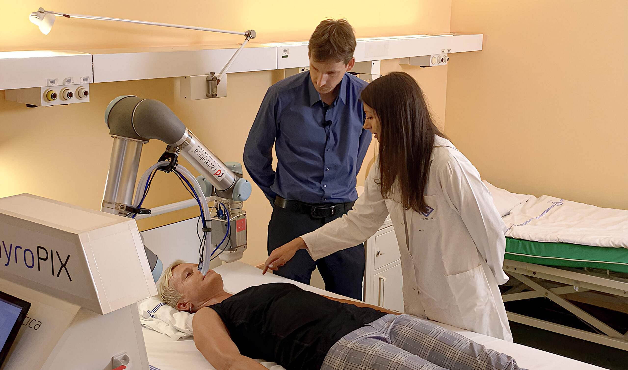
- a collaborative robotic arm enabling both planar and 3D imaging directly at the patient’s bedside. This eliminates the need to transport patients to a dedicated gamma camera suite, thereby reducing the risk of radioactive contamination and radiation exposure of healthcare personnel.
Key Advantages of the ThyroPIX System
- imaging of high radionuclide activities for treatment verification,
- detection of high photon fluxes,
- superior spatial resolution beyond the capabilities of conventional collimator-based gamma cameras,
- mobile design enabling bedside imaging,
- reduced diagnostic activity due to high detection efficiency,
- shorter image acquisition times,
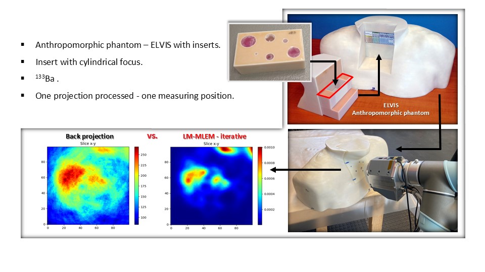
- combined planar and tomographic imaging (SPECT) enabled by the robotic arm.
Software and Hardware Components
The system will include:
- a control unit and software for robotic arm motion control,
- acquisition software for both planar imaging and SPECT,
- image processing and reconstruction software,
- communication modules compatible with NIS/RIS and PACS systems,
- a high-performance workstation with all required software tools.
Additional System Features
The solution will also provide:
- wireless data transfer,
- the possibility of future software upgrades and additional application modules.
The developed system will be designed for:
- diagnostic imaging and monitoring of therapeutic radiopharmaceutical administration,
- high spatial resolution (2D < 2 mm; SPECT < 0.5 ml),
- significantly reduced acquisition times due to the elimination of conventional collimators,
- verification of the spatial distribution of therapeutic radiopharmaceuticals under high photon flux conditions.
Thanks to the flexible collaborative robotic arm, patients can be examined either seated or lying in bed directly in their hospital room, eliminating the need for transportation. This minimizes the risk of radioactive contamination while ensuring precise and reproducible positioning, allowing reliable evaluation of repeated measurements and long-term monitoring of treatment progress.
The project also includes preparation for certification of the medical device as a Class IIa medical device in accordance with the applicable regulatory framework.