




Radalytica a.s. is the project coordinator. The company is responsible for overall project management and for data acquisition using fully digital photon-counting spectral detectors developed by its sister company ADVACAM s.r.o.
Project Objective
To increase the sensitivity of current mammographic examination of surgically resected breast cancer tissue by combining advanced spectral X-ray imaging with artificial intelligence-based data analysis.
Project Concept
The project investigates differences in the spectral responses of biological tissues and evaluates them using artificial intelligence. The objective is to develop an algorithm capable of reliably distinguishing individual tissue types based on their unique spectral characteristics.
Main Project Outcome
The main outcome of the project is a computational algorithm capable of distinguishing malignant tissue from healthy tissue and determining the extent of a lesion directly from spectral data acquired by photon-counting detectors, without the need to reconstruct conventional X-ray images during intermediate processing.

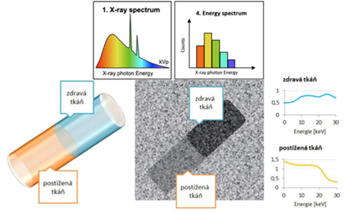

The innovation of the proposed solution lies in the use of a completely new radiographic approach based on hybrid pixel detectors capable of measuring X-ray absorption in predefined energy channels. The acquired spectral data (absorption curves) are subsequently processed using advanced artificial intelligence algorithms.
This approach is unique because it combines state-of-the-art imaging technology with advanced computational methods.
The resulting system will determine the tissue composition of the examined specimen and verify that the identified tissue corresponds to the lesion observed in the X-ray image. This will provide more accurate information about the extent of the lesion and significantly facilitate clinical decision-making by radiologists and oncologists.
In the final phase of the project, the developed methodology will be validated in collaboration with the General University Hospital in Prague, Department of Mammology, using ex vivo X-ray imaging of human breast tumour tissue to verify the research results under clinical conditions.